


How Do You Integrate Ophthalmic Devices with Your EMR and DICOM System?

Your clinic prints exam results, then staff type them into the computer by hand. That takes time, and typos slip into patient records. Many eye clinics still run this way, and the errors scare us.
Integrate your devices with a DICOM-based system and exam images and readings flow straight into patient records. We have seen DICOM integration cut manual data entry errors by 60 to 80 percent in ophthalmology clinics.
The fix is not as hard as it sounds. DICOM is a standard, and modern instruments speak it. Once you understand a few terms, you can plan an integration that pays for itself in staff hours and patient safety.
What is DICOM and why does it matter for modern eye care clinics?
Paper and typing do not scale. When one clinic sees 80 patients a day, staff copy numbers from screens into fields, and mistakes are guaranteed.
DICOM is the global standard for storing and sending medical images. It lets a fundus camera or slit lamp send images, measurements, and patient data to your EMR and PACS, so nothing gets typed twice.
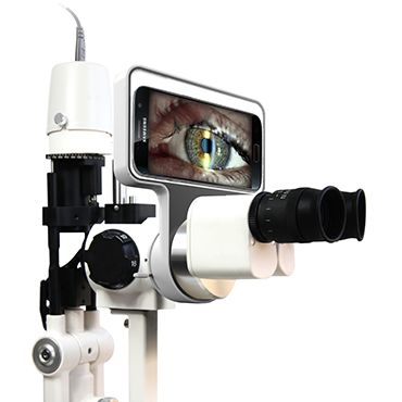
What DICOM actually does
DICOM packages an image with its data. Patient name, exam date, modality, and measurement results travel inside the same file. That means the image and its numbers can never be separated or mixed up, which is exactly what audit and insurance processes demand.
Why eye care clinics need it now
Retinal imaging, OCT scans, and anterior segment photos all produce files that belong in one record. Without DICOM, those files sit on separate machines. With DICOM, a glaucoma patient’s whole history lives in one place, and the next doctor sees it instantly.
The workflow it unlocks
| Task | Without DICOM | With DICOM |
|---|---|---|
| Saving images | Manual export to USB | Automatic to PACS |
| Entering readings | Typed by hand | Sent with the image |
| Finding old exams | Search folders | One patient timeline |
| Sharing with doctors | Email and prints | Secure network transfer |
| Billing support | Rebuilt from paper | Pulled from records |
Start with one device
You do not need to connect everything on day one. Pick the device your clinic uses most, connect it first, and prove the workflow. Once staff see the time saved, they will push for the next connection themselves.
Which ophthalmic instruments commonly support DICOM output today?
Some devices still only export to their own software, so buyers must check before purchase. Most imaging instruments now offer DICOM, but the feature list differs between models.
Fundus cameras, OCT devices, and digital slit lamps commonly support DICOM 3.0 today. Our FC-200 and FC-3100 fundus cameras and our digital slit lamp all send DICOM 3.0 output, so they drop straight into a connected clinic.
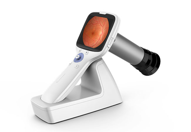
Imaging devices lead the way
Fundus cameras were the first eye care devices to embrace DICOM because retinal photos need precise labeling. OCT followed, since each scan carries dozens of measurements. Digital slit lamp cameras now send anterior segment photos the same way.
Measuring devices and HL7
Pure measuring devices like autorefractors and tonometers often skip DICOM and use HL7 instead. HL7 carries text results rather than images. So a complete clinic uses DICOM for images and HL7 for readings, and the EMR merges both into one patient record.
What our devices support
| Device | Connectivity | What it sends |
|---|---|---|
| Digital slit lamp | DICOM 3.0 | Anterior segment images |
| FC-200 fundus camera | DICOM 3.0 | Retinal images and readings |
| FC-3100 fundus camera | DICOM 3.0 | Retinal images and readings |
| Refraction devices | HL7 options | Refraction results |
Ask for the conformance statement
Every DICOM device should come with a conformance statement. That document lists exactly what the device sends and how. If a supplier cannot give you one, the device probably lacks real DICOM support.
How do you select an EMR system that connects to your diagnostic devices?
An EMR that cannot talk to your devices is just an expensive filing cabinet. Clinics often pick EMR software for billing first, then discover it cannot accept device data.
Choose an EMR with a documented device interface, DICOM storage, and HL7 support. Check that it accepts images from your specific instruments before you sign, and ask for a live demo with your own device data.
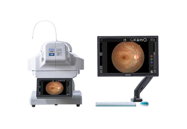
Make a device list first
Write down every instrument you own and every one you plan to buy. Then ask the EMR vendor how each device connects. Vague answers are a warning sign, because the interface is the whole point of the purchase.
Check the interface quality
Some EMRs accept images but bury them in a general folder. You want images attached to the right exam type in the right patient timeline. Test the workflow with a real fundus photo during the demo, and watch where the file lands.
Look at the full workflow
Integration is more than DICOM storage. Orders should reach the device through a worklist, and results should return to the correct encounter automatically. HL7 handles that loop, so ask how the EMR handles order and result messages.
EMR selection checklist
| Requirement | Why it matters |
|---|---|
| DICOM storage | Stores images from cameras and slit lamps |
| HL7 interface | Brings in text readings |
| Device worklist | Pushes patient orders to devices |
| Open API or vendor support | Connects future instruments |
| Per-seat cost model | Keeps scaling affordable |
Test before you commit
Ask the vendor for a trial environment and connect one real device to it. An afternoon of testing beats a year of regret, because integration problems always show up early.
What should you ask your equipment supplier about connectivity before buying?
Most buyers check magnification and light quality but forget connectivity. Then they discover the device cannot feed their system, and the clinic pays for integration work that free DICOM would have done.
Ask about DICOM version, HL7 support, included software, and integration help before you buy. A device with DICOM 3.0 and a conformance statement will save you weeks of setup and thousands in custom work.
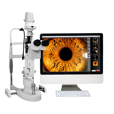
Questions to put to any supplier
Start with the conformance statement, then ask which EMR systems the device has been tested with. Ask whether imaging software is included or sold separately. Ask who configures the network connection and who supports it after installation.
Understand the costs
Some devices need extra software licenses or interface modules before they can talk to your EMR. Ask for those costs in writing before you order. We include imaging and connectivity details in our quotes so clinics can plan the full budget.
Confirm the standard version
DICOM has evolved through versions, and DICOM 3.0 is the modern baseline. Confirm the device sends DICOM 3.0, not an older format or a private file type. Private formats lock your images to one vendor, and that is the opposite of what you want.
Key questions for your supplier
| Question | Why it matters |
|---|---|
| Which DICOM version? | DICOM 3.0 is the modern baseline |
| HL7 support included? | Needed for text readings |
| Software bundled or extra? | Affects total cost |
| Tested with which EMRs? | Predicts setup success |
| Conformance statement available? | Proves real DICOM output |
| Who handles installation? | Defines your support path |
Put it in the contract
Ask for connectivity promises in writing. A supplier who commits to DICOM 3.0 and provides the conformance statement is a partner for the long term, and that partnership is what keeps your records flowing.
Conclusion
Connect your cameras and slit lamps through DICOM 3.0, use HL7 for readings, and pick an EMR that accepts both. Ask suppliers about connectivity before you buy. We at Hongdee include DICOM support and conformance details in every connected device.