


Diabetes is rising everywhere, and eye clinics feel it first. But screening programs fail not from lack of cameras — they fail from ungradable images.
A screening program succeeds when its ungradable image rate stays low. That rate depends on operator training, pupil size protocol, and room lighting — not camera megapixels. Plan the workflow before you buy the camera.
So where do you start? In this article I’ll walk through the equipment a screening lane really needs, why most programs choose non-mydriatic imaging, how to design the capture and grading workflow, what makes a photo gradable, and how to scale from one site to a regional network.
What Equipment Does a Screening Lane Actually Require?
You picture a room full of machines. In practice, a screening lane runs on surprisingly little — if each piece is chosen for the workflow.
A screening lane needs a non-mydriatic fundus camera, a clean desk, controlled lighting, and a data entry point. Add a motorized table and vision chart for full exams, and you can screen hundreds of patients a week.
The Minimum Lane
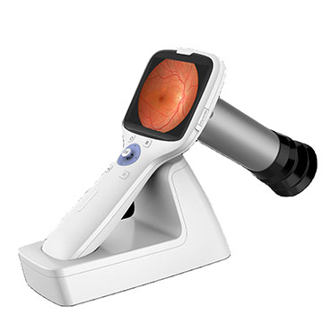
A screening lane starts with four things: a non-mydriatic fundus camera, a dimmable room, a desk with a computer, and a patient register. That is it. The camera captures retinal images, the computer stores them, and the register tracks who was screened and who needs follow-up. Hongdee’s fundus camera range covers this job, including portable models for outreach work in villages and remote clinics.
Lighting matters more than people expect. Non-mydriatic capture works best in a dim room, because bright light shrinks the pupil. Many programs install a simple dimmer switch instead of building a dedicated dark room. That single change can cut your ungradable image rate noticeably.
The Comfortable Lane
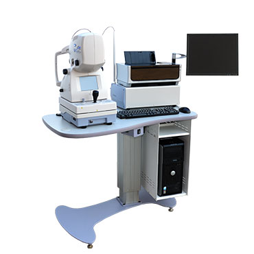
As patient volume grows, add a motorized ophthalmic table and a digital vision chart. Then the same lane can run full exams, not just screening. The motorized table adjusts height for children and wheelchair users, which speeds up patient flow and reduces staff strain.
| Lane type | Core equipment | Extra value |
|---|---|---|
| Minimum | Fundus camera, dim lighting, computer, register | Lowest cost, fastest setup |
| Comfortable | Adds motorized table, vision chart | Full exams in the same room |
| Network hub | Adds grading station, archive server | Central review and reporting |
Choosing Equipment You Can Actually Support
Here is the trap: a program that cannot service its camera dies quietly. Spare parts and calibration wait months, and the lane sits idle. That is why we build our cameras for serviceability and back distributors with a 12-month warranty, free replacement inside the warranty period, and spare parts support. When you choose a supplier, ask about service response time and spare parts availability, not just price.
Why Do Most Programs Choose Non-Mydriatic Imaging?
Dilating every patient slows you down, needs staff, and scares off walk-ins. Non-mydriatic imaging removes that bottleneck.
Most programs choose non-mydriatic imaging because it needs no pupil dilation, no waiting room time, and no extra staff. Patients sit, the operator captures images in dark-room conditions, and the visit ends in minutes.
How Non-Mydriatic Capture Works
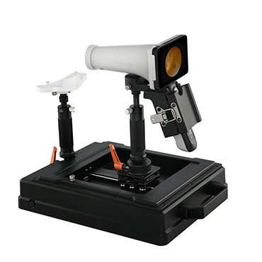
The patient sits at the camera. The room is dim. The operator aligns the eye and captures images in seconds — no drops, no waiting, no dilation consent. This matters in busy settings. A dilated exam ties up a patient for 20 to 30 minutes. Non-mydriatic capture finishes in under five. Multiply that across a hundred patients a week, and the difference is a full day of clinic time.
For the operator, the camera handles much of the work. Autofocus and alignment aids reduce the skill needed to produce usable images. That makes non-mydriatic cameras a realistic choice even where trained ophthalmic staff are scarce — a common situation in rural areas of Africa and Southeast Asia.
When Mydriatic Imaging Still Makes Sense
Dilation is not useless. Some patients have small pupils that never open enough, and some assessments need a wider retinal view. Many programs keep dilation as a second step: capture non-mydriatic first, dilate only when the first image is ungradable.
| Approach | Time per patient | Staff needed | Best fit |
|---|---|---|---|
| Non-mydriatic | Under 5 minutes | One operator | High-volume screening |
| Mydriatic | 20-30 minutes | Operator plus monitoring | Detailed assessment |
The Pupil Size Protocol
Set a simple rule: if the pupil stays small after the room has been dimmed, move the patient to the dilation pathway. Write the rule down, train every operator on it, and review it at monthly meetings. The ungradable rate lives or dies on these small decisions.
How Do You Design the Image Capture and Grading Workflow?
A camera without a workflow is an expensive paperweight. The capture and grading steps decide how many patients you actually process.
Design capture and grading as separate steps: register the patient, capture images, upload, grade, report. One trained operator captures; a grader reads later. This separation keeps throughput high and quality consistent.
The Five-Step Capture Protocol
Every patient follows the same five steps. Step one: register the patient and check the pupil. Step two: dim the room and wait a fixed time for dark adaptation. Step three: capture the image, checking focus and exposure on screen. Step four: upload the image with the patient ID attached. Step five: mark the image as ready for grading.
Each step has an owner. Steps one and two belong to the front desk. Step three belongs to the operator. Steps four and five belong to whoever handles data. When roles are clear, training is fast and errors drop.
Separate Capture from Grading
Grading is a reading task. It needs concentration, a quiet space, and time. If you force the same person to capture and grade, both tasks suffer. Programs that scale well keep the steps separate: operators capture during clinic hours, graders read in batches. This also lets you use remote graders — one specialist can review images from ten sites without travelling.
| Role | Task | Where |
|---|---|---|
| Front desk | Register, check pupil | At the lane |
| Operator | Capture images | At the lane |
| Data clerk | Upload, check labels | At the lane or office |
| Grader | Review, grade, report | Office or remote |
Build the Referral Path Early
A screening program that finds findings but has no referral path creates anxiety, not care. Before you start, agree with a local eye clinic or hospital how referred patients are booked and what information travels with them. This is a workflow step, not a clinical decision, but it decides whether your program earns trust in the community.
What Image Quality Standard Makes a Photo Gradable?
An ungradable photo costs you a patient and a screening slot. Defining “gradable” upfront protects both.
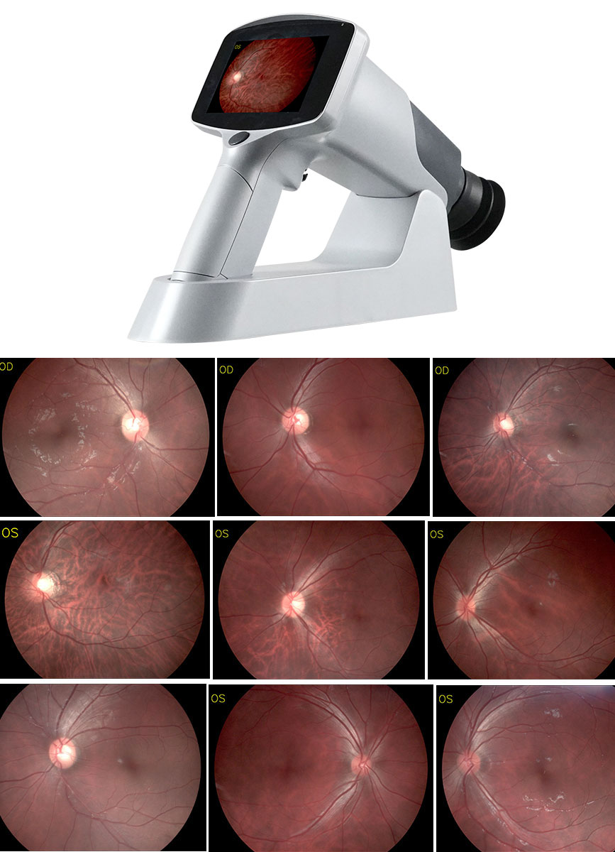
A photo is gradable when the optic disc and macula are visible, focus is sharp, and exposure is even. If any of those fail, the image is retaken on the spot, not filed.

The Gradability Checklist
Train every operator on one checklist. A photo is gradable when the optic disc and macula are visible, the focus is sharp, exposure is even, and the image is labelled with the correct patient. If any item fails, retake the image immediately. Never file a bad image “for now” — it corrupts your data and your reporting.
Why Focus and Exposure Rule
Megapixels grab attention, but they do not fix a blurred image. A sharp, even image from a modest sensor beats a blurry image from a flagship camera every time. Blur comes from patient movement and operator alignment, not from the camera. Exposure problems come from room lighting and flash settings. Both are operator issues, which means both are training issues.
| Quality check | Common cause of failure | Fix |
|---|---|---|
| Disc and macula visible | Wrong alignment | Retake with alignment aid |
| Sharp focus | Patient movement | Re-align, capture again |
| Even exposure | Bright room | Dim lights, adjust flash |
| Correct labelling | Rushed entry | Check ID before upload |
Track the Rate Monthly
Record how many images per hundred are ungradable each month. If the rate climbs, retrain, check the room lighting, and review the protocol. If it falls, you know your training works. This one number tells you more about program health than any equipment specification.
How Do You Scale from One Site to a Regional Network?
One site works. Ten sites fall apart unless data, training, and equipment are standardized from day one.
Scale by standardizing: the same camera model, the same capture protocol, the same grading rules, and one central data review. That way a regional network runs like one big clinic, not ten small experiments.
What Breaks When You Scale
Most programs fail at site two or three. Each new site invents its own habits: different camera settings, different file names, different lighting. Within a year, the central grader cannot compare images across sites. Standardization is the whole game.
Start by choosing one camera model for every site. Spare parts, training, and settings stay identical. Hongdee’s cameras share consistent operation across our range, and our factory capacity — about 5,000 pieces a month — lets distributors stock spare units instead of waiting for repairs. Our 60+ distributors in regions from the Middle East to Latin America keep service close to each site.
| Item | One site | Regional network |
|---|---|---|
| Equipment | Any working camera | Same model everywhere |
| Training | Informal | Certified, repeatable |
| Data | Local spreadsheet | Central review system |
| Grading | Local reader | Central or remote graders |
| Reporting | Optional | Standard monthly report |
The Regional Playbook
Write the playbook before site two opens. It should cover room setup, capture steps, file naming, grading rules, and escalation. Then audit each site against it every quarter. Sites that drift get retrained, not scolded. This is how a network of small clinics behaves like one big one.
Work With Distributors Who Understand Logistics
Shipping cameras across borders raises customs and certification questions. Choose a supplier who has done it before. Hongdee has exported for over 15 years to the USA, Thailand, Singapore, Italy, and Algeria, with CE, FDA, and other certification marks, plus paperwork support for distributors. That experience smooths the customs friction that stalls regional rollouts.
Conclusion
A screening program lives or dies by its ungradable image rate. Standardize equipment, train operators on capture and grading, define gradable images clearly, and scale with one protocol across every site.