


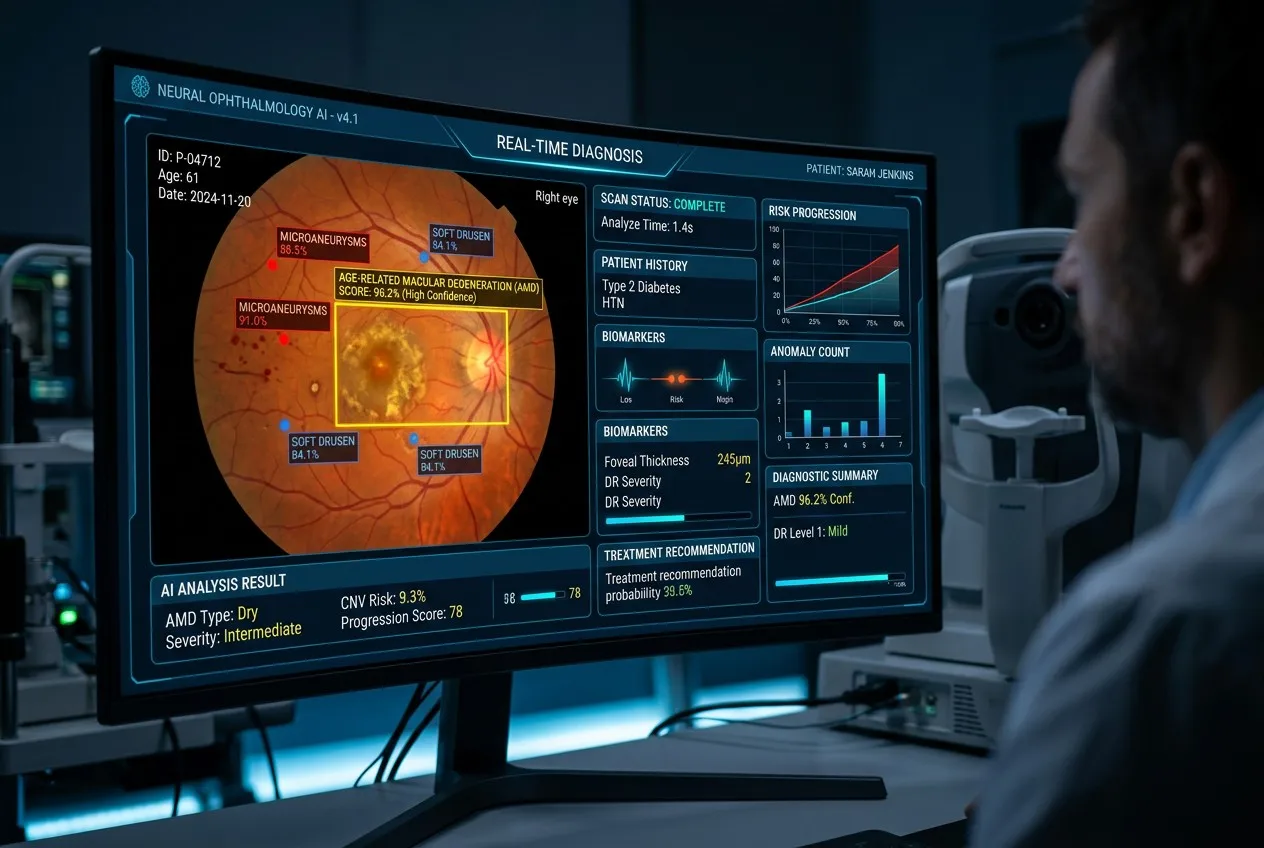
Are you feeling overwhelmed by the rapid speed of AI development in eye care? We see many clinic owners worried about being left behind as technology shifts faster than their budgets.
AI is transforming ophthalmic diagnostics in 2026 by automating routine screenings, improving diagnostic accuracy for retinal diseases, and lowering the barrier for high-quality care in smaller clinics. It allows for faster fundus analysis, enabling practitioners to detect conditions like diabetic retinopathy and glaucoma much earlier than before.
The diagnostic landscape is changing. I have spent 15 years at Hongdee watching equipment evolve from simple manual tools to complex digital systems. Today, the conversation is no longer about the hardware alone. It is about how that hardware speaks to artificial intelligence. Let me walk you through what we are seeing in the industry right now.
What AI Applications Are Already in Ophthalmic Clinics Today?
Many distributors ask if AI is still just a “futuristic concept” or a tool they can sell today. You might feel that your current inventory is already outdated because it lacks “smart” features.
Artificial intelligence is currently used in clinics for automated diabetic retinopathy screening, glaucoma progression analysis, and OCT image segmentation. Platforms like IDx-DR have already gained FDA approval, proving that AI is a clinical reality that helps doctors manage high patient volumes without sacrificing diagnostic quality.

The Rise of Autonomous Screening
In 2026, I see a clear shift toward autonomous systems. These are not just “assistants” anymore. For example, autonomous fundus screening for diabetic retinopathy (DR) is now a standard in many primary care settings. I remember when we first heard about IDx-DR getting FDA clearance. It was a game-changer. Now, similar systems are being integrated into pharmacies and small community clinics. This means eye care is moving closer to the patient.
AI in OCT and Glaucoma Management
It is not just about the fundus camera. Optical Coherence Tomography (OCT) is perhaps the biggest beneficiary of AI. At Hongdee, we track these trends closely. AI algorithms now perform layer segmentation in seconds. This used to take a technician much longer and had a higher margin of error. For glaucoma, AI analyzes the cup-to-disc ratio and compares it to vast databases of healthy and diseased eyes. This predictive modeling helps us see the future of a patient’s vision before they lose it.
Comparison of Current AI Ophthalmic Tools
| AI Tool Type | Primary Function | Clinical Impact |
|---|---|---|
| Fundus Screening | DR & Macular Degeneration | High-speed mass screening |
| OCT Analysis | Layer segmentation & Fluid detection | Precision in surgical planning |
| Biometry AI | IOL power calculation | Improved refractive outcomes |
| Perimetry AI | Visual field progression | Early glaucoma intervention |
How Does AI-Assisted Fundus Screening Actually Work?
Do you wonder if the “black box” of AI is actually reliable enough for your patients? I know many practitioners who fear that a software error could lead to a missed diagnosis.
AI-assisted fundus screening works by using deep learning algorithms to analyze retinal images for specific pathological markers. The software compares the uploaded image against millions of labeled data points, identifying hemorrhages, exudates, and nerve changes with a sensitivity and specificity that often rivals or exceeds human experts.
The Process from Capture to Cloud
When we design fundus cameras at Hongdee, we focus on image clarity. Why? Because AI is only as good as the data it receives. The process is simple: the technician captures a high-resolution image. That image is then sent to a cloud-based AI service. Within seconds, the AI returns a report. It flags the image as “referable” or “non-referable.” This workflow is revolutionary for small clinics that do not have a full-time ophthalmologist on site.
The Power of Low-Cost Cloud Services
I often tell our distributors that you don’t need a $100,000 system to offer AI. Cloud-based AI services have lowered the cost significantly. By using a compatible digital fundus camera—like our Hongdee models—a clinic can pay a small “per-click” fee for AI analysis. This removes the massive upfront investment that used to block small practices from high-end tech. It is a “pay-as-you-go” model for medical excellence.
Key Factors for AI Image Quality
- Resolution: AI needs clear details to see micro-aneurysms.
- Illumination: Uneven lighting can cause “false positives.”
- Field of View: A 45-degree view is standard, but wider is becoming the AI preference.
- Compression: Lossy formats like JPEG can hide subtle signs of disease.
Will AI Replace Ophthalmologists or Just Support Them?
Are you worried that your role as a specialized doctor is being threatened by a computer program? We hear this concern at every trade show, from the AAO in the US to the ESCRS in Europe.
AI will not replace ophthalmologists; instead, it will act as a powerful support tool that handles repetitive screening tasks. By filtering out healthy patients and flagging high-risk cases, AI allows doctors to focus their time and expertise on complex treatments, surgery, and personalized patient care.

Shifting the Burden of Routine Work
I believe the future is about “augmented intelligence.” Think about how much time is wasted looking at healthy retinas. In a typical screening program, 80% of images might be normal. AI can handle that 80% with ease. This leaves the ophthalmologist to deal with the 20% who actually need help. We are not losing jobs; we are gaining efficiency. This is how we will solve the global shortage of eye care professionals.
The Human Element in Medicine
A computer cannot hold a patient’s hand or explain a difficult diagnosis with empathy. AI provides the “what,” but the doctor provides the “why” and the “how.” At Hongdee, when we talk to our 300+ global customers, they all say the same thing: patients want to hear the final word from a human. AI is the most sophisticated tool in your belt, but you are still the craftsman.
Role Evolution in the AI Era
| Task | AI Role | Doctor’s Role |
|---|---|---|
| Screening | High-speed primary filter | Overseeing the screening program |
| Diagnosis | Pattern recognition & grading | Final validation & Clinical context |
| Treatment | Calculating doses or laser paths | Performing surgery & monitoring |
| Patient Care | Data tracking | Counseling & Emotional support |
What Should Distributors Know About Selling AI-Ready Equipment?
Is it getting harder for you to compete on price alone in the crowded ophthalmic market? I know that selling “just another fundus camera” is a race to the bottom that kills your margins.
Distributors should focus on “AI-ready” equipment as a premium offering that provides a higher ROI for clinics. An AI-ready camera is one with high-resolution digital output, stable API connectivity, and a track record of compatibility with leading AI diagnostic platforms like Eyenuk or Google Health.
Creating a Premium Value Proposition
I always tell our partners: don’t sell a box, sell a solution. When you sell an AI-ready Hongdee fundus camera, you are selling the ability for the clinic to generate more revenue through screening services. This is a much stronger argument than “our price is 10% lower.” AI readiness creates a “moat” around your business. Your customers will rely on you for the integration, not just the hardware.
The Importance of Open Systems
Some big brands try to lock users into their own expensive AI ecosystems. I think this is a mistake. The best AI-ready equipment is “platform-agnostic.” This means the images can be exported to any AI service the doctor chooses. At Hongdee, we ensure our digital outputs follow international standards. This flexibility is a huge selling point for distributors who want to offer their clients the best of both worlds.
Checklist for AI-Ready Hardware
- [ ] Minimum 12MP resolution: Essential for detail.
- [ ] DICOM Compatibility: The standard for medical imaging.
- [ ] External API Support: Can it talk to third-party software?
- [ ] Non-Mydriatic Capability: Needed for high-speed screening.
- [ ] Ease of Use: Can a technician (not just a doctor) take perfect images?
How Do I Prepare My Clinic for the AI Diagnostic Era?
Do you feel like your clinic’s infrastructure is stuck in the 1990s while the world moves to 2026? It is frustrating to want the best technology but feel limited by your current setup.
Preparing your clinic for AI involves upgrading to high-quality digital imaging hardware, ensuring a robust high-speed internet connection for cloud processing, and training your staff on standardized image capture protocols. You must also consider the legal and data privacy aspects of sharing patient data with AI providers.

Hardware First, Software Second
I have seen clinics try to buy AI software before they have a camera that can take a decent photo. It doesn’t work. Your first step is getting a reliable, high-resolution fundus camera or OCT. We have helped many clinics transition by providing equipment that fits their budget today but is ready for the AI of tomorrow. Our experience as a Topcon OEM for 3 years taught us that precision in hardware is the foundation of everything else.
Training and Workflow Integration
AI is not a “plug and play” magic wand. You need to change how your staff works. Who takes the photos? How are they uploaded? How does the doctor review the AI report? We suggest starting small. Use AI for one type of screening—like diabetic retinopathy—and build your workflow around it. Once that is smooth, expand to glaucoma or macular degeneration. This step-by-step approach prevents burnout and ensures the tech actually gets used.
The Three Pillars of AI Readiness
| Pillar | Focus Area | Action Step |
|---|---|---|
| Infrastructure | Internet & Data Security | Upgrade to fiber & HIPAA/GDPR storage |
| Equipment | Digital Output Quality | Replace old manual/film units with Hongdee digital |
| Knowledge | Staff training | Certify technicians in standardized imaging |
Conclusion
The AI diagnostic era is here to stay, offering massive opportunities for efficiency and better patient outcomes. By choosing AI-ready equipment and focusing on staff training, you can future-proof your clinic and provide world-class eye care starting today.