


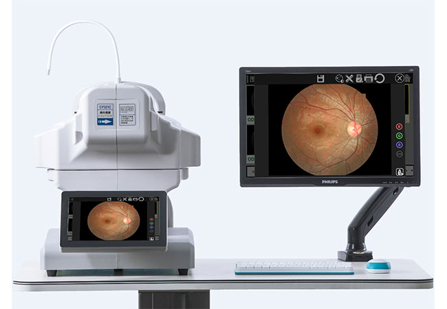
Adding cataract screening is not the same as building a cataract surgery theatre. The first expansion must define what the clinic can screen, document, and refer.
Start with a referral-ready pre-op screening pathway: history, visual function, examination, relevant imaging, and documented escalation. Add complete biometry only when qualified staff, equipment, quality control, and the surgical team support it.
At Hongdee, we separate initial screening from a complete surgical work-up. Equipment helps organise information. It does not make a patient a surgical candidate or replace an ophthalmologist’s assessment.
What belongs in an initial cataract pre-op screening pathway?
A cataract referral can fail when a clinic collects a photograph but misses visual function, history, examination, or urgency. A camera is not a pathway.
Initial screening should support a qualified professional’s history and examination, visual acuity, refraction where appropriate, anterior-segment assessment, relevant ocular health checks, and referral documentation. The exact scope depends on local practice and clinical governance.

Build around the referral question
We ask the clinic to define what the receiving surgeon needs to know. Is the goal to identify reduced vision that may be cataract-related? To document the anterior segment? To identify a possible urgent condition? To prioritise a referral appointment? Each question changes the equipment and record requirements.
A slit lamp can support a professional anterior-segment examination. A visual acuity chart supports a functional measure. Refraction equipment may help distinguish correctable refractive error from persistent reduction, within the operator’s scope. A fundus camera may support selected documentation or referral, but a normal-looking image does not rule out every condition and an image must be interpreted appropriately.
We also recommend a structured referral form. Record patient identity, symptoms and history, visual acuity, relevant measurements, examination findings, images if taken, clinician name, date, urgency, and receiving service. Specify what remains unknown. This keeps the screening service honest and gives the surgical team useful context.
| Screening element | Possible equipment category | Boundary |
|---|---|---|
| Visual function | Vision chart or refraction tools | Does not identify every cause of loss |
| Anterior segment | Slit lamp | Requires trained examination |
| Documentation | Fundus camera where indicated | Image is not a complete diagnosis |
| Referral record | Secure record system and form | Needs governance and follow-through |
Set a safe stop point
The clinic should know when to stop screening and refer urgently. Red flags, poor image quality, unexplained visual loss, and examination findings outside the service scope should not be forced into a routine cataract label. Qualified professionals define the clinical criteria.
When do you need complete pre-operative biometry rather than screening tools?
Pre-op screening and surgical planning are often blended in buyer conversations. They are different services with different quality requirements.
Complete pre-operative biometry generally requires measurements such as axial length and corneal parameters, plus clinician review and an appropriate calculation workflow. A screening camera, slit lamp, or ordinary refractor is not a substitute for a biometer.
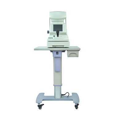
Define the handoff before buying a biometer
If a clinic only identifies and refers possible cataract cases, the surgical centre may perform biometry. If the clinic will provide a complete pre-operative work-up, it needs the surgical team’s specification for measurements, formulas, data transfer, repeat rules, and quality review. We recommend obtaining that specification before purchasing an instrument.
Axial length is not produced by an ordinary auto refractor. Corneal curvature or keratometry is also a distinct measurement. A biometer may combine functions, but buyers must verify the exact model and configuration. We do not infer that any product shown in this article performs biometry, and we do not claim that Hongdee’s pictured equipment covers every cataract planning need.
Ask who repeats a questionable measurement, who reviews outliers, how dense cataracts or unusual eyes are handled, and what happens when optical acquisition fails. Some cases may require another measurement method or specialist review. Those are professional and model-specific decisions.
Treat calculation as a controlled clinical step
IOL calculation is not just a button on a machine. The surgical team chooses the clinical method and validates the workflow. The record should preserve the measurement inputs, eye, date, operator, formula or calculation setting where applicable, lens information, and reviewer. If data are exported, confirm the format and responsibility for transcription.
For equipment procurement, this means a brochure claiming “cataract solution” is not enough. Request the manual, measurement specifications, supported workflow, software details, training plan, and service route. Keep screening and surgical-planning claims separate in marketing and staff instructions.
How should you select screening equipment without buying an entire surgical unit?
Expansion often starts with a narrow service. Buying every device at once can create unused hardware, training gaps, and unclear responsibility.
Choose equipment in stages: first support reliable screening and referral, then add biometry or imaging only when demand, staffing, surgical partnerships, records, and quality controls justify the next step.
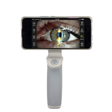
Use a staged package
Stage one may include visual acuity, appropriate refraction resources, a slit lamp, documentation, and a referral relationship. Depending on the pathway, a fundus camera may add useful images. Stage two may add a biometer when a qualified team is responsible for complete pre-operative measurements. Stage three may address operating-room or surgical equipment, which is outside an initial screening expansion and needs a separate plan.
This staging prevents a common category error. A clinic can offer high-quality screening without claiming to offer surgical planning. It can also avoid paying for a specialist instrument before it has a trained operator and a receiving team that agrees on the data format.
| Expansion stage | Main question | Procurement focus |
|---|---|---|
| Screening | Can we identify and refer appropriately? | Basic examination and records |
| Referral support | Will images improve the handoff? | Image quality, privacy, review route |
| Complete pre-op work-up | Does the surgical team accept our data? | Biometry, keratometry, workflow, QA |
| Surgery | Are we equipped and authorised to operate? | Separate clinical and regulatory project |
Plan space, people, and records
A slit lamp needs a suitable examination position and trained operator. Imaging needs privacy, storage, transfer, and review. Biometry needs fixation, repeatability, and a clinician-led quality process. Each addition expands the service manual and maintenance file.
Use Hongdee’s digital slit lamp and ophthalmic motorized table pages to frame questions about equipment categories. Confirm the exact model and intended use before representing any device as suitable for a cataract pathway.
What acceptance and referral controls protect the new service?
A new screening service is vulnerable at the handoff. Missing accessories, poor images, unclear urgency, and undocumented limits can delay the next decision.
Use model-specific acceptance tests, image and examination quality checks, a written referral form, staff competency records, privacy controls, downtime steps, and a review process for rejected or incomplete referrals.
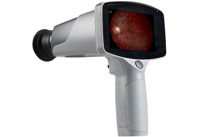
Accept the workflow, not only the box
Before delivery, list the model, accessories, software, electrical requirements, documents, and training scope. At receipt, record identity and damage. After installation, test the agreed functions according to the manual. For a camera, check image capture, storage, export, and privacy controls. For a slit lamp, check illumination, movement, optics, and positioning controls as applicable. These are operational checks, not evidence of universal clinical accuracy.
Run a supervised referral simulation. Can staff record the patient correctly? Can they identify a poor image? Can the receiving service open the file? Is urgency visible? Does the patient know where to go next? If any answer is no, quarantine the affected workflow and correct it before routine use.
Keep claims within the service boundary
We recommend wording such as “cataract referral screening” when that is the actual service. Avoid saying “complete pre-op assessment” unless the clinic has the agreed measurements, professional oversight, and surgical-team acceptance. Avoid implying that a fundus image, refractor, or slit lamp alone establishes surgical suitability.
The WHO Package of Eye Care Interventions provides a useful service-planning context. The American Academy of Ophthalmology cataract clinical resources can inform professional review, while local rules govern practice. For procurement principles, see the WHO procurement process guide. Sources checked 2026-10-12; this article is procurement education, not patient treatment advice.
Conclusion
Expand in stages: screen and refer first, then add complete biometry only with the right team and workflow. At Hongdee, we help buyers specify equipment without overstating what screening proves. Request a model-specific quote.