



Are you still relying on hand-drawn sketches to document your patients’ eye conditions? If you aren’t capturing high-definition photos and videos of the anterior segment, you are missing out on better patient education and more accurate medical records.
A digital slit lamp imaging system transforms a standard diagnostic tool into a powerful documentation and education device. It allows you to capture, store, and share high-resolution images of the eye, which is essential for tracking disease progression, securing insurance reimbursements, and enabling remote consultations.
At Hongdee, we have been at the forefront of this digital shift. With three years of OEM experience for major brands and a global network of 60+ distributors, we’ve seen how “going digital” changes a clinic’s reputation. In this guide, we will explore why digital imaging is no longer a luxury but a core requirement for any modern ophthalmic practice.
What Does a Digital Slit Lamp Imaging System Do That a Traditional Slit Lamp Cannot?
A traditional slit lamp is for “seeing.” A digital system is for “remembering” and “sharing.” This distinction is the foundation of modern medical documentation.
Unlike traditional units, a digital slit lamp imaging system uses a high-performance camera module (integrated or external) to record what the doctor sees through the oculars. It creates a permanent visual record that can be analyzed frame-by-frame, compared over time, and sent to other specialists via EMR systems.
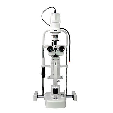
From Sketches to 4K Video
In the old days, a doctor would draw a “clock face” diagram to show a corneal ulcer or a cataract. But sketches are subjective. A “large” ulcer to one doctor might be “medium” to another. A digital system captures the exact color, depth, and size of the lesion. Our 300+ customers often tell us that having 4K video of a moving eye is far more valuable than a single static photo for detecting subtle issues like dry eye or pupillary responses.
Background Illumination and Filters
Digital systems often come with specialized lighting. You get background illumination that lights up the whole eye while the “slit” stays focused on the pathology. This creates professional-grade photos that look like they came from a textbook. We ensure our Hongdee imaging modules include yellow filters (for better cobalt blue fluorescein shots) and diffusers to reduce glare, which is a major problem in amateur eye photography.
Real-Time Patient Viewing
With a digital system, you can “mirror” the image to a large monitor. This means the patient sees what you see. When they see the red vessels of their own eye or the cloudiness of their cataract on a 24-inch screen, their “buy-in” for your treatment plan increases dramatically. This is a “marketing” power that traditional slit lamps simply don’t have.
| Feature | Traditional Slit Lamp | Digital Slit Lamp System |
|---|---|---|
| Documentation | Hand-drawn / Mental notes | HD Photo / 4K Video |
| Objectivity | Subjective (Observer bias) | Objective (Visual proof) |
| Patient Education | Verbal description only | Real-time visual display |
| Collaboration | Hard to share | Instant export (JPG/MP4) |
| Workflow | Faster per patient | Adds 1-2 mins for capture |
What Are the 4 Real Benefits Clinics Get From Upgrading?
We talk to distributors in Brazil, Italy, and the UAE every day. They tell us that “better photos” is only the beginning. The real benefits are about the “3 Rs”: Revenue, Referrals, and Reliability.
The four primary benefits include enhanced patient education (increasing procedure conversion), superior tracking of chronic diseases (like glaucoma or cataracts), simplified insurance billing with visual proof, and the ability to participate in tele-ophthalmology networks for remote expert opinions.
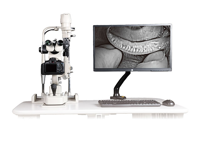
1. Increased Treatment Compliance
When a patient sees their “dry eye” film breaking up on screen, they are much more likely to buy the expensive eye drops or agree to a procedure. Visual proof removes the “sales” feeling and replaces it with “medical necessity.” We’ve seen clinics in Singapore and the US increase their conversion rates for elective procedures by 20% after installing a digital imaging system.
2. Precise Disease Monitoring
How much has that pterygium grown in 6 months? If you have two photos side-by-side, you know the answer. Without them, you are guessing. This is critical for medical-legal protection too. If a patient claims their condition worsened under your care, your digital history is your best defense. This “reliability” is why hospital procurement teams prioritize digital systems.
3. Faster Insurance Approvals
Insurance companies are getting stricter. They want proof before they pay for a surgery. A high-quality image of a “grade 3” cataract or a corneal scar makes the approval process much faster. This reduces the administrative burden on your staff and improves your clinic’s cash flow.
4. Tele-Ophthalmology and Referrals
We are seeing a massive global trend toward remote consultations. If you are a primary care optometrist in Australia or Algeria, you can send an image to a surgeon in the city for an instant second opinion. This keeps the patient in your care “loop” and ensures they get the right treatment faster. Digital imaging is the “ticket” to joining these modern healthcare networks.
| Benefit | Impact on Practice | Value Level |
|---|---|---|
| Conversion | Higher revenue from procedures | High |
| Documentation | Reduced medical-legal risk | Critical |
| Referrals | Better connection with specialists | Moderate |
| Billing | Fewer rejected insurance claims | High |
Smartphone-Based vs. Integrated Camera Modules: Which One Fits Your Budget?
This is a common question from our 60+ distributors. There is a huge price gap between a “clip-on” and a professional imaging workstation.
Smartphone-based adapters are affordable and portable, using the phone’s high-quality camera for quick capture. Integrated camera modules (workstations) offer higher clinical precision, better lighting control, and seamless DICOM integration with EMR systems, making them the better choice for busy, professional clinics.
Smartphone Adapters: The Entry Point
If you are a startup in California or a mobile clinic in Thailand, a smartphone adapter is a great start. They cost very little and use the 12MP-48MP camera already in your pocket. However, they have downsides. The lighting is often uneven, and you have to struggle with “phone focus” vs. “slit lamp focus.” Also, storing medical photos on a personal phone can be a HIPAA or GDPR privacy risk.
Professional Integrated Modules
These are what we specialize in at Hongdee. These modules (like our Phonto or Phoenix series) sit between the oculars and the objective lens. They use professional CMOS sensors designed for low-light medical imaging. They have a “split-beam” design so you can see through the oculars while the camera captures the image. The software is built for doctors, not for Instagram, ensuring all photos are tagged with the patient ID and stored securely.
ROI Comparison
While the integrated module costs more upfront, it saves time. With a smartphone, you have to align the phone, take the shot, and then email it to yourself. With a professional module, you press a “joystick button,” and the photo is in the patient’s file instantly. In a clinic seeing 20+ patients a day, the “time saved” pays for the professional module in less than a year.
| Factor | Smartphone Adapter | Integrated Workstation |
|---|---|---|
| Price | Very Low | Moderate to High |
| Image Quality | Good (Depends on Phone) | Excellent (Medical Grade) |
| Workflow Speed | Slow (Manual Transfer) | Fast (Auto-Sync) |
| Privacy/Security | Low (Personal Device) | High (Encrypted EMR) |
| Durability | Low (Plastic Parts) | High (Metal Housing) |
Can You Retrofit a Digital Imaging System to Your Existing Slit Lamp?
You don’t always need to buy a brand-new slit lamp to go digital. This is a secret that big-name manufacturers don’t want you to know.
Yes, you can retrofit most major slit lamp brands with a digital imaging module. Hongdee’s Phonto and Phoenix modules are specifically designed to be compatible with brands like Marco, Topcon, Zeiss, Haag Streit, Keeler, and Nidek, allowing you to upgrade your current equipment for a fraction of the cost.

Compatibility is Key
Many doctors think: “I have a 10-year-old Topcon; I have to throw it away to get digital photos.” That is false. As long as your slit lamp has a standard “beam splitter” port or a compatible “eyepiece” mount, we can likely digitalize it. We have helped 300+ customers globally save thousands of dollars by providing the right “bridge” or “adapter” for their existing optics.
The Retrofit Process
Installing a module like our Phoenix system takes about 15 minutes.
1. You remove the binocular head.
2. You insert the beam splitter/camera module.
3. You re-attach the head.
4. You plug the USB into your PC.
Our technical team provides remote support to distributors in Europe and the Middle East to ensure the “back-focal length” is perfect, so the image on the screen is as sharp as the image in your eyes.
Why Retrofit?
- Sustainability: Keep using the high-quality glass optics you already own.
- Cost-Efficiency: Spend 70% less than buying a new “All-in-One” digital slit lamp.
- Familiarity: You don’t have to learn a new machine’s mechanical controls; only the software is new.
- Resale Value: A digitalized older slit lamp is much easier to sell if you ever decide to retire or move.
How Is Digital Imaging Changing Referral Patterns and Remote Consultations?
The world is getting smaller. Digital imaging is the “internet” of eye care, connecting local shops to global experts.
Digital imaging allows for “Tele-Ophthalmology,” where high-resolution files are sent across the world for instant analysis. This allows primary care providers to manage more complex cases locally with the support of remote specialists, reducing travel time for patients and increasing the scope of local practices.

The Rise of the “Hub and Spoke” Model
We are seeing many optical chains in the UK and Australia use this model. The “spokes” are small shops with digital slit lamps. They capture images of anything suspicious and send them to the “hub” — a central hospital where a senior surgeon reviews 100 cases a day. This is incredibly efficient. It ensures that only the patients who truly need surgery actually travel to the hospital.
AI Integration
The next step, which we are already seeing in China and Singapore, is AI screening. The digital slit lamp sends the image to an AI server. Within seconds, the AI flags signs of “dry eye severity” or “corneal neovascularization.” This acts as a “second set of eyes” for the doctor. To participate in this future, you must have a system that produces “clean,” high-resolution images that AI can read.
Global Connectivity
At Hongdee, we ship to 60+ countries. We’ve seen doctors in Algeria consulting with colleagues in Italy using our Phonto modules. This global knowledge sharing improves patient outcomes everywhere. Whether you are a distributor looking for the next big trend or a clinic owner wanting to stay relevant, digital imaging is the bridge to the future of eye care.
Future-Proofing Your Practice
- Step 1: Get a professional imaging module (Phonto/Phoenix).
- Step 2: Integrate it with a DICOM-capable EMR.
- Step 3: Join a referral or tele-medicine network.
- Step 4: Explore AI diagnostic plugins as they become available.
Conclusion
Digital slit lamp imaging is no longer optional for those who want to provide top-tier eye care. It improves education, ensures better records, and connects you to the global tele-medicine network. Upgrade your existing lamp today to protect your practice’s future.