
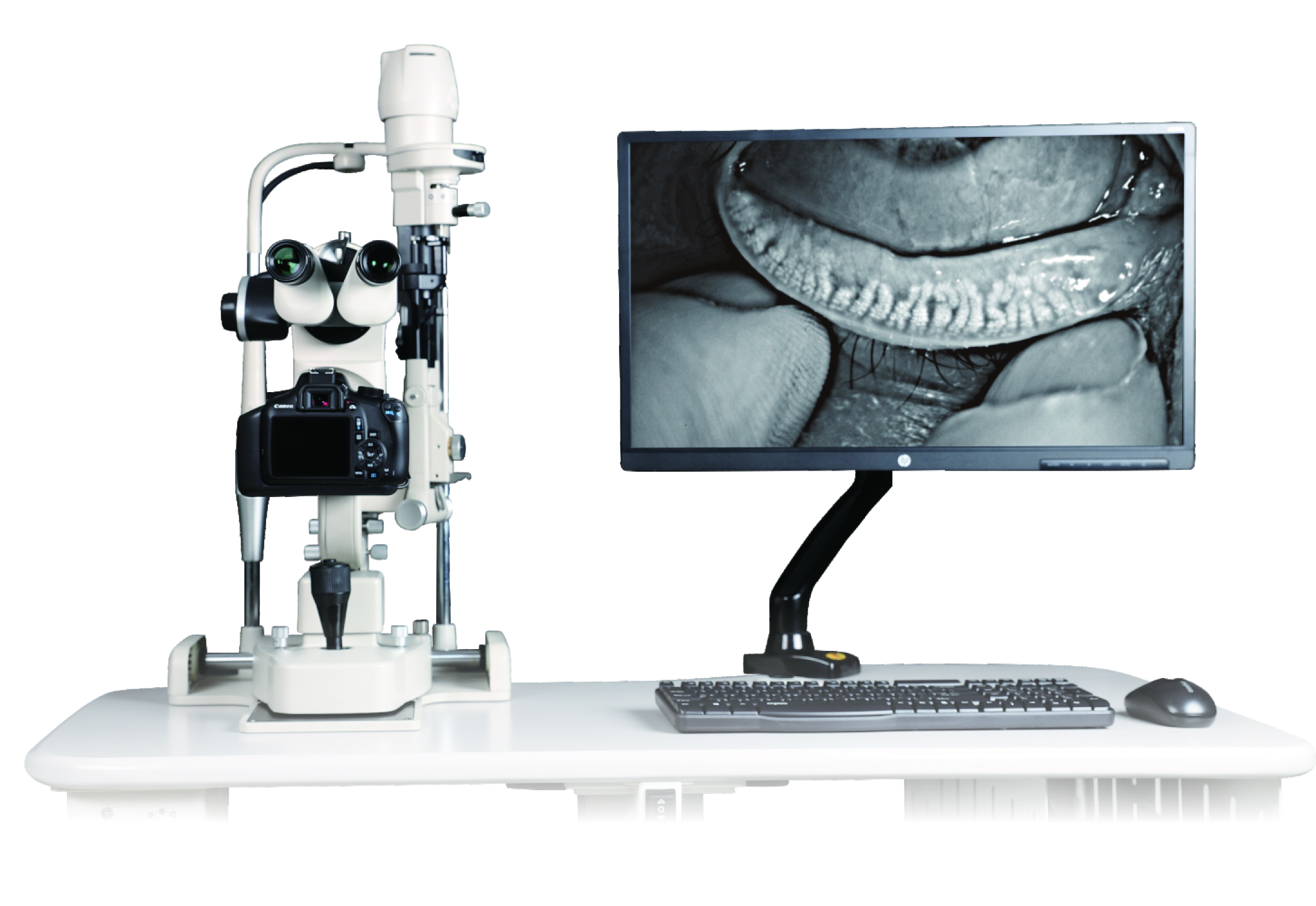
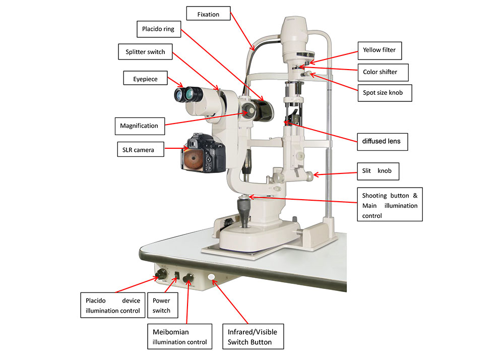
In Hongdee, there are multiple slit lamp types for eye diagnosis, like cornea, cortical cataract, dry eye, or glaucoma. Aupha V D, specially designed for a dry eye examination, is exquisite and precise, with no loss of nasal side ring image. A large aperture ensures images’ high definition and color rendition. Connected to an optical system with hidden wire, more controllable and stable.
How To Use Slit Lamp For Dry Eye Exam?
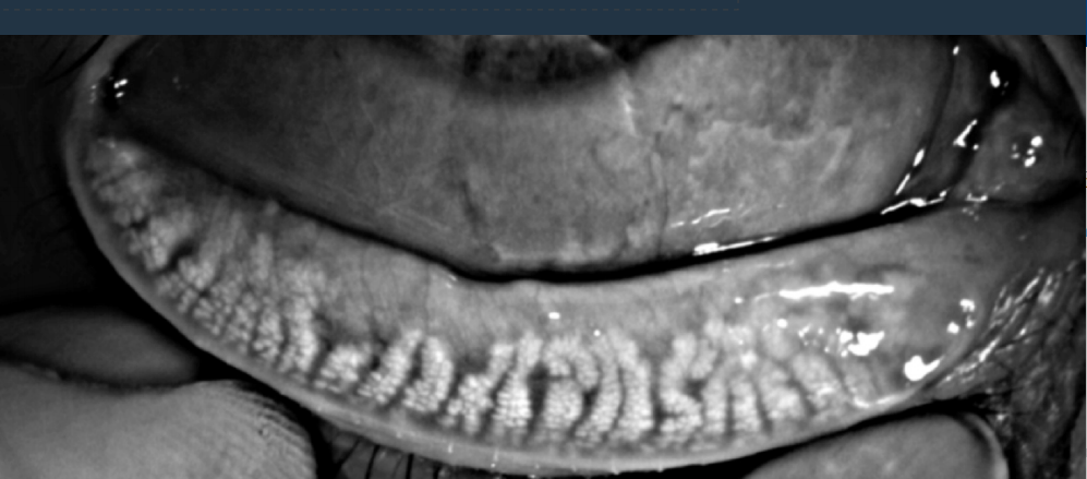
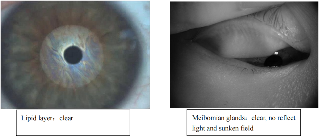
Meibomian gland function evaluation
Clear observation of meibomian glands Intuitively diagnose MGD
Quantitative analysis of meibomian gland deletion
With an infrared imaging system and HD digital camera, the area of meibomian gland deletion can be intuitive present. Meibomian gland loss severity can be objectively evaluated by comparing with the contrast models of meibomian gland deletion level.
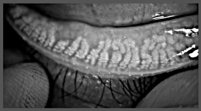
Multistep magnification observation of meibomian gland
Multistep optical magnification systems support lossless image magnification. Meibomian gland distribution can be observed in the images of lower magnification, the area of missing glands can be clearly present. The detail of gland acini can be observed in the images of higher magnification, the outline is clear. It supports the clinicians in the timely treatment of the meibomian gland lesions, and diagnosis and treatment efficiency is improved.
Meibomian gland dysfunction(MGD) is the main cause of lipid layer deficiency, which leads to evaporative dry eye. It is characterized by obstruction of gland ducts, falling off the glands, or qualitative change and quantitative change of gland secretions. MGD results in tear film fast evaporation and hyperosmolarity, ocular surface inflammation, epithelium damage, and discomfort. Infrared imaging is a fast and comfortable method of meibomian gland examination, which is the only non-invasive technology for observing meibomian glands in a clinic.
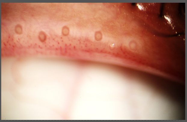
Eyelid margin analysis
Eyelid margin change is one of the typical characteristics of MGD, including eyelid margin morphologic change and meibomian gland opening change. It hasmultistepopticalmagnificationsystem, meeting the observation requirement of eyelid margin morphologic change and meibomian gland opening change. Meibomian gland and gland opening distribution can be observed in the images of lower magnification, and the details of the eyelid margin and gland opening change can be observed in the images of higher magnification. The images provide the intuitive diagnosis basis for eyelid margin abnormality or MGD.
Lipid layer analysis
It analyzes the thickness, quality, and quantity of the lipid layer by comparing it with graded contrast models, and diagnoses MGD combined with meibomian gland and eyelid margin analysis. A specially designed tear film imaging unit enlarges the aperture and reduces the light loss, increasing the image resolution and color rendition. The lipid layer morphology and coating process can be dynamically observed by HD videos, which have exquisite frames and strong hierarchical effects.
Dry eye disease comprehensive examination
All-round examination items Comprehensive dry eye evaluation
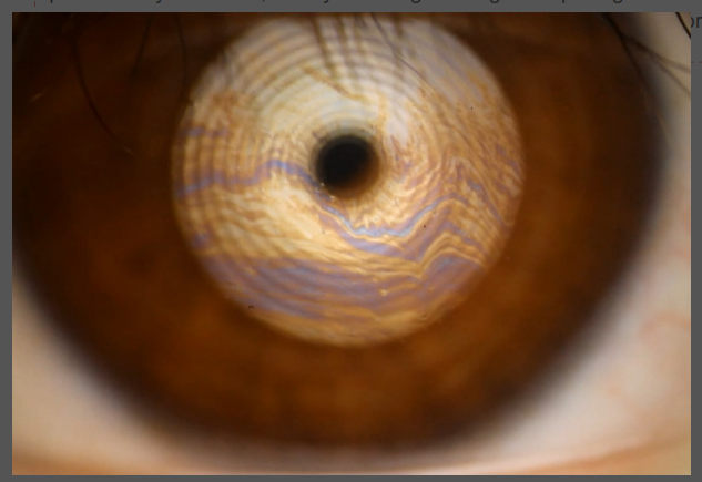
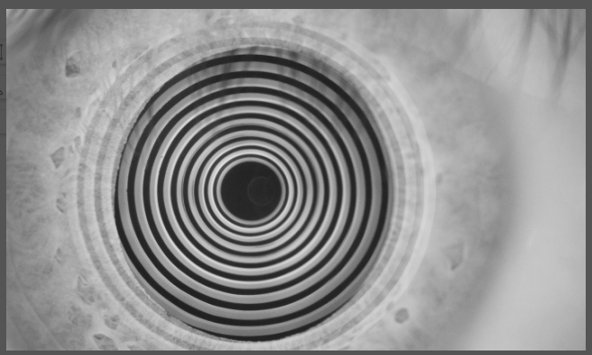
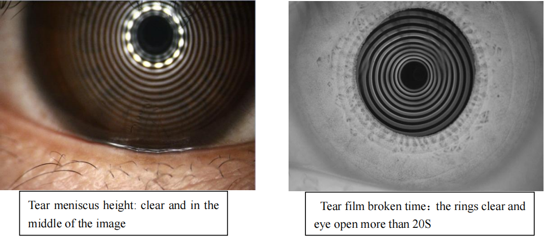
Non-invasive breakup time ( NIBUT)
Dry eye disease (DED) is characterized by tear film instability. SLM-KD3 quantitatively evaluates the state of tear film stability by identifying the ring image changes on the tear film. Tear film imaging units utilize infrared light, which is non-invasive and non-stimulative. It directly faces the eye being examined, the ring image is complete and the nasal side ring image has no defect, which ensures the complete video record. Compare with fluorescein staining, non-invasive examination provides a more comfortable experience for the patients, an automatic timing system is more accurate and efficient, and the operation process is simplified.
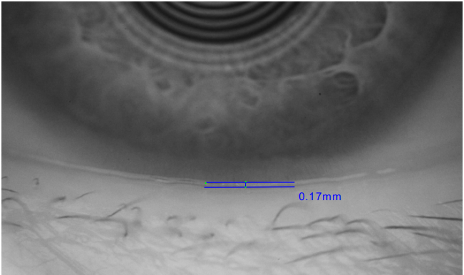
Tear meniscus height (TMH)
Compare with the Schirmer test, SLM-6E(A) shorten the examination time by more than 10 times, and the result is more accurate. Tear film imaging units utilize infrared light, which is non-invasive and non-stimulative. A specially designed tear film imaging unit enlarges the aperture and reduces the light loss, increasing the image resolution. Multistep optical magnification system support image optical magnification for more detailed observation of tear meniscus morphology. If the tear meniscus is discontinuous or uneven, conjunctivochalasis (dry eye complication) or palpebral margin abnormal (MGD) can be simultaneously examined.
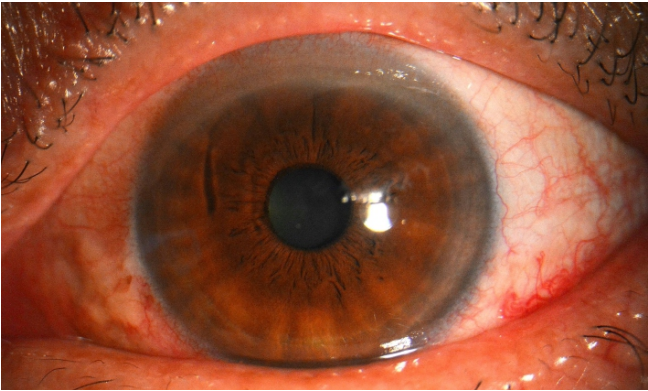
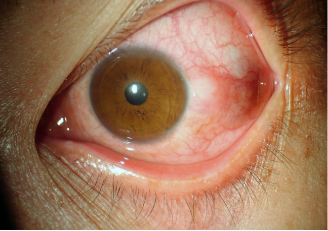
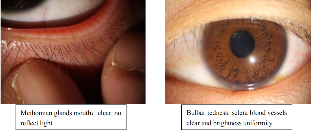
Bulbar redness scan (R-scan)
Common bulbar redness includes conjunctival congestion, ciliary congestion, and mixed congestion, which is probably caused by dry eye disease or conjunctive. In the dry eye examination, bulbar redness is preferentially considered to be aqueous deficient dry eye (ADDE). SLM-6E(A) captures images in high definition, automatically grades the conjunctival congestion and ciliary congestion, and evaluates the severity of bulbar redness.
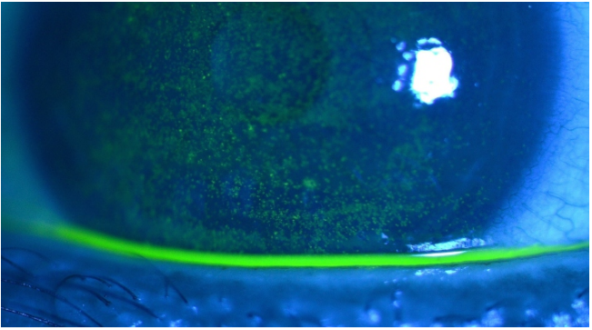
Corneal staining
Assess the corneal epithelial completeness, and check mucous deficient dry eye or corneal inflammation. Unique optical magnification systems support lossless image magnification. Five blue filter combinations enhance the effect of fluorescein staining, cornea details can be observed clearly, which spares no details of the small corneal lesion.
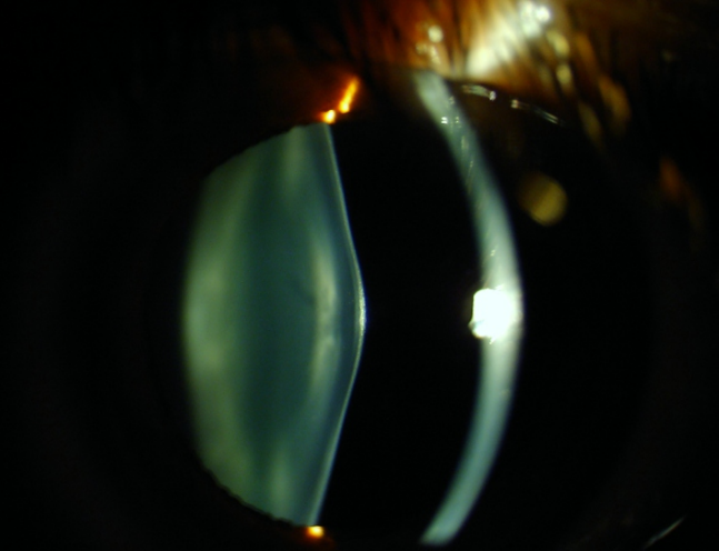
Anterior segment examination on the same device
More practical function More efficient examination
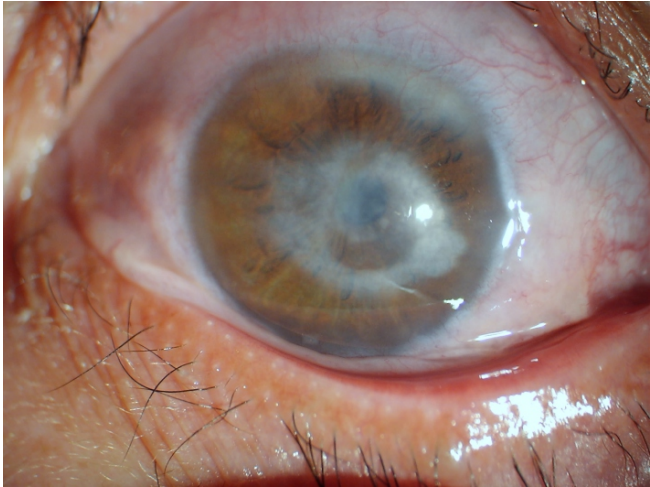
Dry eye complications examination
Without changing the device, dry eye complications can be rapidly checked, including cornea damage (cornea scarification, cornea inflammation, cornea ulcer, cornea perforation), corneal nebula, conjunctivochalasis, which improves the checking efficiency and patient satisfaction. Binocular eyepiece for better observation with a stereoscopic view. Multistep optical magnification systems support lossless image magnification. Built-in contrast-enhanced filter for more clear observation of lesion detail.
Anterior segment examination
All anterior segment examinations can be done on the same device, more efficient. Anterior segment examinations include but are not limited to Ocular surface disease, measurement of cornea diameter and pupil diameter and lesion area, corneal lesions, lens lesions, and 1/3 forepart of vitreous lesions. Examinations of the fundus and posterior segment of the vitreous can be done with other additional accessories, corneal contact lens matching examination for identifying the contact lens contraindications.
Exam Images
Finally, dry eye slit lamp automatically generates reports according to the examination results without manually filling them in. The illustrated report is easy to understand, the operation process is simpler. The visible and intuitive reports facilitate communication with patients.
If buying an upper-level standard dry eye slit lamp for ophthalmologists, please send a formal inquiry for a quick quote. Hongdee will be your perimeter business partner.