

In advanced eye care, standard surface-level observations are no longer the benchmark for comprehensive diagnostics. Optical Coherence Tomography (OCT) has transitioned from a high-end luxury to an absolute clinical necessity. Often described as an “optical ultrasound,” an OCT system utilizes near-infrared light waves to capture micrometer-resolution, cross-sectional images of the ocular tissue.
For modern eye clinics and hospitals, implementing an OCT platform is the single most effective way to detect asymptomatic pathologies, manage chronic retinal diseases, and elevate the overall standard of patient care.
1. Unmatched Sub-Surface Visualization (The Ocular Ultrasound)
Traditional diagnostic instruments, such as standard fundus cameras or digital slit lamps, only provide a two-dimensional view of the retina’s surface. However, the most critical blinding pathologies develop hidden beneath the surface layer.
An OCT scan penetrates deep into the tissue structure, mapping individual histological layers in real time.
-
Micron-Level Resolution: It measures the precise thickness of the Retinal Nerve Fiber Layer (RNFL) and the ganglion cell complex.
-
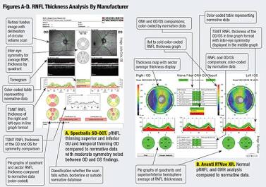
Early Glaucoma Detection: As shown in clinical topography reports, it cross-references a patient’s nerve fiber thickness against a normative database. This color-coded mapping allows clinicians to detect localized thinning—indicative of early glaucoma—years before the patient experiences actual visual field loss.
-
Identifying Structural Anomalies: It reveals microscopic structural defects such as sub-retinal fluid accumulation, macular holes, and choroidal neovascularization (CNV) that are completely invisible to the naked eye.
2. Definitive Diagnosis and Management of Macular Diseases
Age-Related Macular Degeneration (AMD) and Diabetic Retinopathy are leading causes of irreversible blindness worldwide. Effective intervention relies heavily on the timing of the diagnosis. An OCT system fundamentally transforms how these conditions are managed:
| Clinical Phase | Traditional Method (Fundus Exam) | Advanced Method (OCT System) |
| Detection | Identifies advanced structural changes like large hemorrhages or severe exudates. | Detects micro-vacuoles of fluid and early drusen beneath the retinal pigment epithelium (RPE). |
| Classification | Difficult to differentiate between “wet” and “dry” AMD without invasive dye injections. | Differentiates fluid type immediately via non-invasive cross-sectional structural scans. |
| Treatment Tracking | Relies on subjective visual acuity changes and gross structural observations. | Provides quantitative, micron-level tracking of central macular thickness to measure anti-VEGF injection efficacy. |
By utilizing quantitative measurements, ophthalmologists can monitor exactly how many microns of fluid have decreased following a treatment session, allowing for a highly personalized and objective therapeutic approach.
3. Streamlining Surgical Workflows and Pre-Operative Planning
An OCT system is just as crucial for anterior segment specialists and refractive surgeons as it is for retinal experts. Modern platforms offer specialized lenses to scan the front structures of the eye, drastically improving surgical predictability.
Pre-Op Scan ──► Precise Micro-Measurements ──► Custom Surgical Plan ──► Post-Op Verification
-
Premium IOL Power Calculations: For cataract surgeries, anterior OCT imaging maps the exact corneal epithelial thickness and anterior chamber depth, ensuring the selection of the perfect premium intraocular lens (IOL).
-
Glaucoma Shunt Placement: It provides high-resolution visualization of the iridocorneal angle, allowing surgeons to plan and verify the placement of Micro-Invasive Glaucoma Surgery (MIGS) devices.
-
LASIK/PRK Safety: Refractive specialists use it to measure the residual stromal bed thickness pre- and post-laser ablation, mitigating the risk of corneal ectasia (thinning and bulging).
Conclusion: The Backbone of Modern Evidence-Based Ophthalmic Care
Investing in an OCT system is no longer just about adopting new technology—it is about establishing clinical certainty. By replacing subjective visual assessments with definitive, quantifiable, sub-surface data, the OCT platform empowers eye clinics to preserve patient sight through early detection and precise treatment monitoring. For any practice aiming to deliver advanced, competitive, and truly evidence-based eye care, the OCT system is an indispensable asset.